Home
Team
Research
Publications
Research Funding and Networks
News
Join Us
Team
Research
Publications
Research Funding and Networks
News
Join Us
合成所
深圳先进院
中国科学院
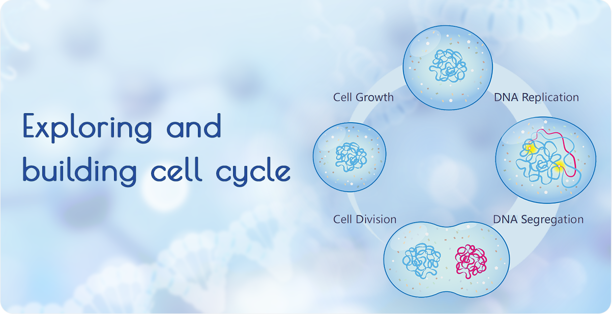
Cell Cycle
SynCell
Bacterial Cancer Therapy