IEEE TMI | High Time Resolution CT Imaging
Recently, researchers Li Yinsheng and Liang Dong from the Medical Artificial Intelligence Research Center of Shenzhen Institute of Advanced Technology, Chinese Academy of Sciences, and the Key Laboratory of Medical Imaging Science and Technology System (National), in cooperation with Shanghai Lianying Medical Technology Co., Ltd. and Beijing Tiantan Hospital affiliated to Capital Medical University, have published their research results of high time resolution CT imaging in the top journal of international medical imaging IEEE Transactions on Medical Imaging (IEEE TMI, IF=10.6). Researcher Li Yinsheng is the first author and corresponding author of the paper, while Researcher Liang Dong is the main corresponding author of the paper.
The reconstruction of fault images requires the scanned object to be static within the minimum complete data acquisition time. When the requirement is not met, the reconstructed image has a time averaged error (i.e., low temporal resolution). When the rack speed is fixed, using shorter viewing angle range data to reconstruct the image, that is, improving the temporal resolution of the reconstruction method, can reduce this error. However, using shorter viewing angle range data to reconstruct images violates the data completeness condition, resulting in incomplete data errors (as shown in Figure 1). Existing methods, including traditional FBP and finite angle reconstruction methods, cannot minimize both types of errors simultaneously.
Unlike using finite angle reconstruction to improve temporal resolution, this paper proposes a method that achieves the goal of improving temporal resolution through extrapolation along the time dimension. Specifically, a single time point image reconstructed from complete data is extrapolated along the time dimension to obtain multiple time point images, each corresponding to a higher temporal resolution. However, due to the existence of an infinite number of extrapolation rules, there are an infinite number of extrapolation results. Without appropriate constraints, it is impossible to find the correct result from it. Therefore, it is technically very difficult to extrapolate a single time point image to obtain accurate multiple time point images.
During the CT data acquisition process, the dynamic information of the scanned object is sequentially recorded in each perspective projection data. The time required to obtain projection data from a single perspective is very short, with high time resolution, such as 0.1 milliseconds for diagnostic CT or 25 milliseconds for C-arm cone beam CT. We naturally thought of using a single perspective projection data to constrain the aforementioned extrapolation process along the time dimension. By minimizing the inconsistency between the extrapolated image sequence and the measured projection data, the optimal extrapolation rule is obtained.
To achieve the above idea, this paper proposes a deep temporal dimension extrapolation method with data consistency constraints, which utilizes data consistency constraints to learn the optimal extrapolation rules to improve temporal resolution (as shown in Figure 2). The innovation of the proposed method lies in: (1) increasing the CT temporal resolution to the system limit (i.e. the time required to obtain single view projection data -0.1 milliseconds for diagnostic CT or 25 milliseconds for C-arm cone beam CT), thereby minimizing both time averaged error and incomplete data error; (2) This method is suitable for single short scan data acquisition protocols and non sparse imaging tasks; (3) This method explicitly uses projection data measured by each patient, so that the learned extrapolation rules are optimal for each patient (as shown in Figure 3).
Shenzhen Advanced Technology Research Institute of the Chinese Academy of Sciences has recently been approved as the Key Laboratory of Medical Imaging Science and Technology System (National). The laboratory is committed to achieving international leadership in principle innovation and technological breakthroughs in new generation imaging systems and advanced medical diagnosis and treatment. Intelligent medical imaging is a key research direction supported by laboratories. This research work is one of the series of achievements achieved with the support of the laboratory. This research work has received support from the National Natural Science Foundation of China Youth Science Fund and the Shenzhen Basic Research Key Project.
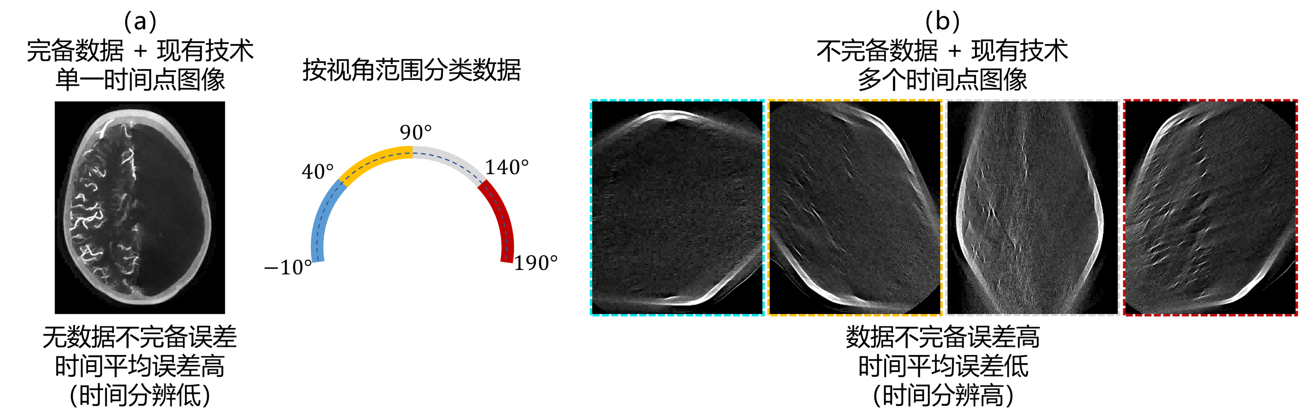
图1. (a)完备数据使用现有技术仅能重建单个时间点图像,该图像无数据不完备误差,但时间平均误差高,时间分辨低;(b)按视角范围分类数据,每个数据子集不完备,使用现有技术重建多个时间点图像,每个图像时间平均误差低,时间分辨高,但不完备误差高。
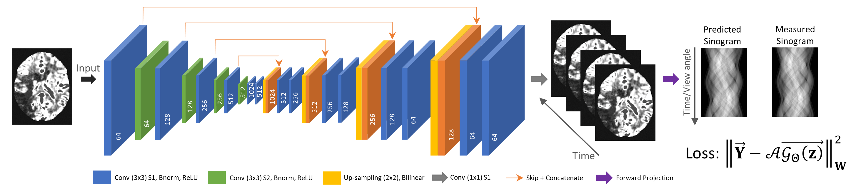
图2. 方法流程图。
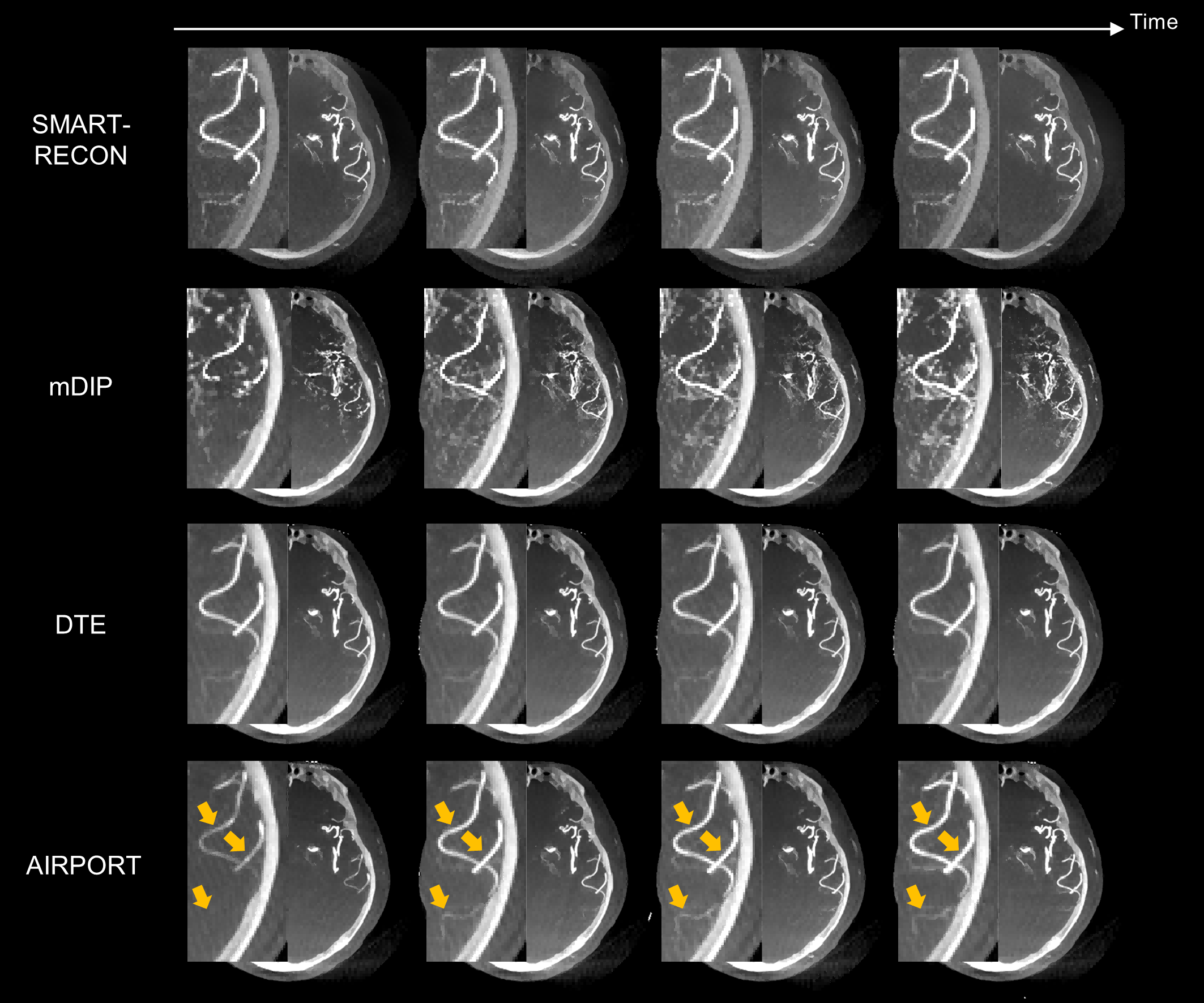
图3. 人体实验数据结果。如黄色箭头所示,提出的AIRPORT方法能够正确地重建由造影剂注射导致的动脉强度值的变化。